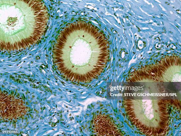
Epididymis, light micrograph - stock photo
Epididymis, light micrograph. The lumen of the duct (white) is lined with pseudostratified epithelium (green), which is made up of columnar cells with elongated nuclei and rounded basal cells with circular nuclei. The brown fibres extending into the lumen are stereocilia. The duct is surrounded by a layer of smooth muscle (blue). Magnification: x120 when printed at 10 centimetres wide.

Get this image in a variety of framing options at Photos.com.
PURCHASE A LICENCE
All Royalty-Free licences include global use rights, comprehensive protection, and simple pricing with volume discounts available
$500.00
+GST NZD
Getty ImagesEpididymis Light Micrograph High-Res Stock Photo Download premium, authentic Epididymis, light micrograph stock photos from Getty Images. Explore similar high-resolution stock photos in our expansive visual catalogue.Product #:99310965
 Download premium, authentic Epididymis, light micrograph stock photos from Getty Images. Explore similar high-resolution stock photos in our expansive visual catalogue.Product #:99310965
$500+GST$50+GST
Getty Images
In stockDETAILS
Credit:
Creative #:
99310965
Licence type:
Collection:
Science Photo Library
Max file size:
4842 x 3638 px (41.00 x 30.80 cm) - 300 dpi - 4 MB
Upload date:
Release info:
No release required
Categories: